
Tubercular Spinal Infection: What is it?
Overview:
Infectious spondylitis used to be primarily caused by tuberculosis. The only choices for treating paralysis prior to the development of effective chemotherapy were time and surgery. Currently, 2% to 3% of all documented instances of M. tuberculosis are caused by tubercular bone and joint infections. Between one-third and one-half of bone and joint infections are caused by spinal tubercular infections. The most often affected region is the thoracolumbar spine. Males and females are nearly equally infected, although the rate of infection appears to rise with age.
The reason behind the infection:
Acid-fast-positive, caseating granulomas, with or without pus, are the pathological hallmark of the infection. When Langerhans-type giant cells are present, microscopic analysis usually reveals tubercles made of monocytes and epithelioid cells that form tiny masses with central caseation. Abscesses contain necrotic debris and grow by taking the easiest route. Skin sinuses develop, empty, and mend on their own.
A bone’s response to an infection might range from severe to nonexistent. The infection spreads beneath the anterior and posterior longitudinal ligaments of the spine, avoiding the intervertebral discs. Permanent brain damage is more likely to occur from an epidural infection.
Spinal TB signs and indicators
The early stages of the disease are characterized by slowly progressing constitutional symptoms, such as fever, weight loss, weakness, malaise, and night sweats. A late indication of paralysis and bone collapse is pain. Hoarseness can result from cervical involvement due to respiratory stridor (also called Millar asthma), dysphagia, and recurring paralysis of the laryngeal nerve. The development of an anterior abscess in the neck may cause these symptoms. Cervical illness has been known to cause sudden death following erosion into the major arteries.
STORIES OF PATIENT SUCCESS
Neurological symptoms may grow and wane and typically appear late. Rectal tone and motor function are reliable indicators of prognosis. According to Jain et al., a 76% encroachment on a CT scan can be accommodated by the spinal canal without causing neurological abnormalities. Researchers found that in an outdoor hospital, 60% to 90% of patients with Pott paraplegia recovered after extended bed rest.
How to make a diagnosis:
Chronic disease is suggested by laboratory investigations. Anemia, hypoproteinemia, and a little increase in ESR are among the findings. Skin testing is not diagnostic, although it might be useful. Due to the possibility of skin slough from a severe reaction, the test is not recommended for patients who have previously had a tuberculous infection. It is also not helpful for patients who may be experiencing a reactivation of the disease.
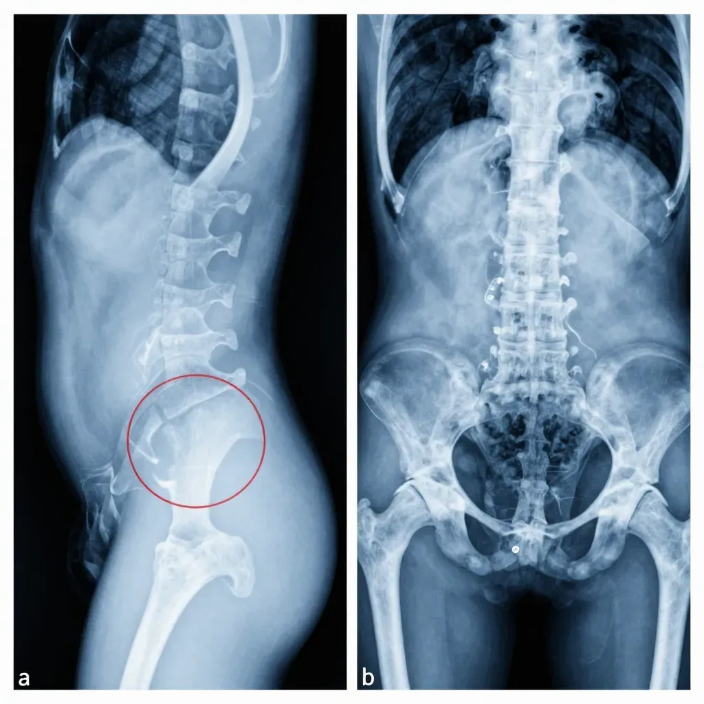
Localized osteopenia and a slight reduction in one or more disc spaces are Early radiography findings. Later discoveries include vertebral collapse, which Seddon dubbed “concertina collapse” due to its accordion-like characteristics. Radiographic findings of soft-tissue edema and its subsequent calcification are very predictable. With or without contrast, CT scanning makes it possible to assess the disease process and the extent of neurological impairment more accurately. MRI makes it possible to further define the disease process. The only MRI findings that helped differentiate spinal tuberculosis from neoplasia were the presence of bone pieces and the formation of abscesses. However, none of these tests can prove the presence of tuberculosis. Researchers found that patients with disseminated TB benefited most from 67 Ga scanning.
A biopsy of the lesion is necessary for a definitive diagnosis, which is based on the organism’s culture. Percutaneous methods that are controlled by CT or radiography are typically sufficient. 89% of cases had epithelioid granulomas, 83% had positive acid-fast bacilli cultures, and 52% had positive acid-fast bacilli smears. If a needle biopsy is risky or ineffective, or if other open procedures are necessary, an open biopsy can be necessary.
Missed and delayed diagnoses are frequent. Primary bone tumors (such as osteosarcoma, chondrosarcoma, myeloma, eosinophilic granuloma, and aneurysmal bone cyst), sarcoidosis, giant cell tumors of bone, pyogenic and fungal infections, secondary metastatic disease, and bone deformities like Scheuermann disease are examples of differential diagnoses.
The toxicity of the chemotherapeutic drugs and the duration of treatment necessitate a definitive diagnosis by culture of a biopsy material. Following surgery, none of the patients experienced paraplegia.
Better outcomes when radical surgery is carried out with chemotherapeutic treatment in terms of deformity, recurrence, paralysis development, and resolution. Surgical intervention was not necessary to resolve paraplegia. Whether or not cast immobilization was used, prolonged bed rest did not work. The preferred course of treatment in cases where facilities for radical surgery are not accessible is ambulatory chemotherapy.
Which treatments are available for treating spinal tuberculosis?
Spine TB / स्पाइन टीबी में सर्जरी कब करनी चाहिए
There is a wide range of justifications for surgery when neurological symptoms are absent. The danger of kyphosis and collapse is greatly increased when many vertebrae are involved. The most straightforward method for these individuals may be open biopsy for diagnosis, débridement, and grafting with or without anterior instrumentation. Radical surgical treatment is also indicated in cases of recurrence of the disease and resistance to chemotherapy.
Indications for surgery in early or late disease include signs and symptoms of cord compression, gradual impairment of pulmonary function, advancement of the kyphotic deformity, and severe kyphosis with active disease. Heart and respiratory failure are the main reasons why surgery is not recommended.
After anterior decompression and grafting, Posterior fusion—with or without spinal instrumentation—is recommended to avoid late collapse and stress fracture of the graft in cases where more than two vertebrae are involved and anterior instrumentation is not utilized. Rarely is posterior fusion by itself recommended at this time.
Posterior fusion alone has been associated with high failure rates and late advancement of kyphotic deformity, with or without fatigue fracture of the fusion. As long as it is sufficiently long, the tricortical iliac crest is the recommended bone transplant material for all levels. Every time débridement and grafting are done, external immobilization is required.
Following cervical and cervicothoracic surgeries, halo (vest, cast, or pelvic) immobilization is employed for three months. Following thoracic and thoracolumbar surgeries, either detachable or nonremovable thoracolumbar immobilization is utilized until the grafts have fully healed, which can take anywhere from nine to twelve months. Following low lumbar surgeries, lumbosacropelvic immobilization is employed. It should be applied from the hip to the knee of at least one leg for 6 to 8 weeks. Thoracolumbosacral immobilization is then applied until the infection has cleared up and the graft has healed.